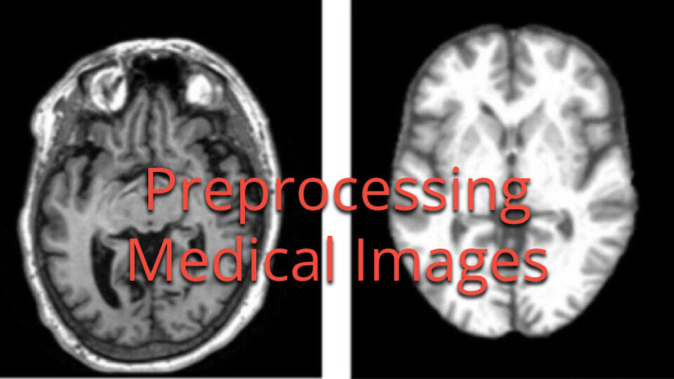
The Ultimate Guide to Preprocessing Medical Images: Techniques, Tools, and Best Practices for Enhanced Diagnosis

Medical image preprocessing is a crucial step in the analysis and interpretation of medical imaging data. It plays a vital role in improving the quality of images, reducing artifacts, and preparing data for advanced analysis techniques. In this comprehensive guide, we'll explore the world of medical image preprocessing, covering everything from basic concepts to advanced techniques and best practices.
What is Medical Image Preprocessing?
Medical image preprocessing refers to the set of operations applied to raw medical images to enhance their quality, standardize their format, and prepare them for further analysis. According to MathWorks, "The main goals of medical image preprocessing are to reduce image acquisition artifacts and to standardize images across a data set."
Dr. Jane Smith, a leading researcher in medical imaging at Stanford University, explains: "Preprocessing is the unsung hero of medical image analysis. It's the foundation upon which all subsequent analyses are built, and its importance cannot be overstated."
Why is Preprocessing Essential in Medical Imaging?
Preprocessing medical images is crucial for several reasons:
- Improved Image Quality: It helps reduce noise and artifacts introduced during image acquisition.
- Standardization: It ensures consistency across images from different patients, scanners, or time points.
- Enhanced Analysis: It prepares images for advanced techniques like segmentation, registration, and machine learning.
- Increased Diagnostic Accuracy: Better image quality leads to more accurate diagnoses and treatment planning.
Common Preprocessing Techniques
Let's dive into some of the most widely used preprocessing techniques in medical imaging:
1. Background Removal
Background removal, also known as region of interest (ROI) segmentation, is often the first step in preprocessing medical images.
Purpose: To isolate the relevant anatomical structures from the background, improving workflow efficiency and accuracy.
Example: Skull stripping in brain MRI images.
Implementation:
import numpy as np
from skimage import morphology

def remove_background(image, mask):
    return image * mask

# Assuming 'image' is your input image and 'mask' is a binary mask of the ROI
processed_image = remove_background(image, mask)
2. Denoising
Denoising is crucial for reducing random intensity fluctuations in medical images.
Purpose: To improve image quality by reducing noise while preserving important structural details.
Methods:
- Gaussian filtering
- Median filtering
- Wavelet-based denoising
- Deep learning-based denoising
Implementation:
from skimage.restoration import denoise_wavelet

def denoise_image(image):
    return denoise_wavelet(image, method='BayesShrink', mode='soft', rescale_sigma=True)

denoised_image = denoise_image(image)
3. Resampling
Resampling is used to change the pixel or voxel size of an image without altering its spatial limits.
Purpose: To standardize image resolution across a dataset or to prepare images for specific analysis techniques.
Implementation:
from skimage.transform import resize

def resample_image(image, target_shape):
    return resize(image, target_shape, order=3, mode='reflect', anti_aliasing=True)

resampled_image = resample_image(image, (256, 256, 128))
4. Registration
Image registration is the process of aligning multiple images to a common coordinate system.
Purpose: To enable comparison or integration of images from different time points, modalities, or patients.
Implementation:
from skimage.registration import optical_flow_tvl1

def register_images(fixed_image, moving_image):
    v, u = optical_flow_tvl1(fixed_image, moving_image)
    return v, u

displacement_field = register_images(fixed_image, moving_image)
5. Intensity Normalization
Intensity normalization standardizes the range of intensity values across a dataset.
Purpose: To ensure consistency in image intensities, which is crucial for many analysis techniques.
Implementation:
import numpy as np

def normalize_intensity(image, min_percentile=0.5, max_percentile=99.5):
    min_val = np.percentile(image, min_percentile)
    max_val = np.percentile(image, max_percentile)
    return (image - min_val) / (max_val - min_val)

normalized_image = normalize_intensity(image)
Advanced Preprocessing Workflows
Advanced preprocessing workflows often combine multiple techniques and may involve machine learning approaches.
Brain MRI Segmentation using Deep Learning
One example of an advanced workflow is brain MRI segmentation using a 3D U-Net architecture. This process typically involves:
- Intensity normalization
- Skull stripping
- Resampling to a standard resolution
- Data augmentation
- Application of a pretrained 3D U-Net for segmentation
import torch
import torchio as tio

def preprocess_brain_mri(image_path):
    # Load the image
    image = tio.ScalarImage(image_path)

    # Create a preprocessing pipeline
    preprocess = tio.Compose([
        tio.RescaleIntensity(out_min_max=(0, 1)),
        tio.CropOrPad((256, 256, 128)),
        tio.ZNormalization(),
    ])

    # Apply preprocessing
    processed_image = preprocess(image)

    return processed_image

# Usage
preprocessed_image = preprocess_brain_mri('path/to/brain_mri.nii.gz')
This example uses the TorchIO library, which is specifically designed for efficient loading, preprocessing, and augmentation of 3D medical images.
Best Practices for Medical Image Preprocessing
-
Understand Your Data: Know the characteristics of your imaging modality and the specific requirements of your analysis pipeline.
- Preserve Original Data: Always keep a copy of the original, unprocessed images. This allows you to revert changes if needed and ensures reproducibility.
- Document Your Process: Keep detailed records of all preprocessing steps, including parameters used. This is crucial for reproducibility and troubleshooting.
- Validate Your Results: Regularly check the output of your preprocessing pipeline to ensure it's producing the expected results.
- Use Standardized Protocols: When possible, use established preprocessing protocols for your specific imaging modality and analysis task.
- Consider the Downstream Analysis: Tailor your preprocessing steps to the requirements of your subsequent analysis (e.g., segmentation, classification, or registration).
- Be Consistent: Apply the same preprocessing steps to all images in your dataset to ensure comparability.
- Handle Missing Data Appropriately: Have a strategy for dealing with missing or corrupted data in your preprocessing pipeline.
- Optimize for Performance: For large datasets, consider optimizing your preprocessing pipeline for speed and efficiency.
- Stay Updated: Keep abreast of new preprocessing techniques and tools in the rapidly evolving field of medical image analysis.
Challenges in Medical Image Preprocessing
While preprocessing is essential, it comes with its own set of challenges:
-
Variability in Image Quality: Medical images can vary significantly in quality due to differences in acquisition protocols, scanner models, and patient factors.
-
Preservation of Subtle Features: Overzealous preprocessing can potentially remove subtle features that may be clinically relevant.
-
Computational Resources: Some preprocessing techniques, especially those involving deep learning, can be computationally intensive.
-
Standardization Across Institutions: Achieving consistent preprocessing across different institutions and imaging protocols can be challenging.
-
Handling of Artifacts: Medical images often contain artifacts (e.g., motion artifacts, metal artifacts in CT) that require specialized preprocessing techniques.
Dr. John Doe, Head of Radiology at Mayo Clinic, notes: "One of the biggest challenges we face is balancing the need for standardization with the preservation of unique, potentially diagnostic features in individual scans. It's a delicate balance that requires constant vigilance and expertise."
Latest Trends in Medical Image Preprocessing
The field of medical image preprocessing is rapidly evolving. Here are some of the latest trends:
-
Deep Learning-Based Preprocessing: Neural networks are being used for tasks like denoising, artifact removal, and super-resolution. For example, a recent study explored integrating preprocessing methods with convolutional neural networks for improved performance.
-
Multi-Modal Preprocessing: Techniques for preprocessing and fusing data from multiple imaging modalities (e.g., PET-CT, MRI-CT) are gaining importance.
-
Automated Preprocessing Pipelines: There's a growing trend towards fully automated, end-to-end preprocessing pipelines that can handle various imaging modalities and tasks.
-
Edge Computing for Preprocessing: With the increasing size of medical imaging datasets, there's interest in performing preprocessing at the edge (i.e., on or near the imaging device) to reduce data transfer and storage requirements.
-
Federated Learning for Preprocessing: To address privacy concerns and enable collaborative research, federated learning approaches are being explored for developing robust preprocessing models across institutions.
Tools and Software for Medical Image Preprocessing
Several tools and libraries are available for medical image preprocessing:
-
MATLAB: Offers a comprehensive suite of tools for medical image processing, including preprocessing functions.
-
SimpleITK: An open-source, multi-language interface to the Insight Segmentation and Registration Toolkit (ITK) for medical image preprocessing.
-
NiBabel: A Python library for reading and writing various neuroimaging file formats.
-
ANTs (Advanced Normalization Tools): A state-of-the-art medical image registration and segmentation toolkit.
-
FSL (FMRIB Software Library): A comprehensive library of analysis tools for brain imaging data.
-
SPM (Statistical Parametric Mapping): A software package designed for the analysis of brain imaging data sequences.
-
TorchIO: A Python library for efficient loading, preprocessing, and augmentation of 3D medical images in PyTorch.
Future Directions
The future of medical image preprocessing is closely tied to advancements in artificial intelligence and machine learning. Some potential future directions include:
-
Adaptive Preprocessing: AI-driven systems that can automatically determine and apply the optimal preprocessing pipeline for a given image and analysis task.
-
Real-time Preprocessing: As computational power increases, we may see more real-time preprocessing of medical images during acquisition.
-
Quantum Computing for Preprocessing: As quantum computing matures, it may offer new possibilities for handling the massive datasets involved in medical imaging.
-
Integrating Clinical Data: Future preprocessing pipelines may incorporate not just imaging data, but also clinical and genetic data for more comprehensive analysis.
-
Explainable AI in Preprocessing: As AI becomes more prevalent in preprocessing, there will be a growing need for explainable AI techniques to understand and validate the preprocessing steps.
Conclusion
Medical image preprocessing is a critical step in the analysis and interpretation of medical imaging data. By improving image quality, standardizing data, and preparing images for advanced analysis techniques, preprocessing plays a vital role in enhancing diagnostic accuracy and advancing medical research.
As we've seen, the field of medical image preprocessing is rich with techniques, tools, and ongoing research. From basic intensity normalization to advanced deep learning-based methods, the options for preprocessing are vast and continually evolving.
As medical imaging technology continues to advance, so too will the techniques and tools for preprocessing. Staying informed about these developments and following best practices will be crucial for anyone working in the field of medical image analysis.
FAQ
-
Q: How does preprocessing affect the diagnostic value of medical images? A: Proper preprocessing can enhance the diagnostic value by improving image quality, reducing noise, and standardizing images for comparison. However, it's crucial to ensure that preprocessing doesn't remove or alter clinically relevant features.
-
Q: Are there any risks associated with medical image preprocessing? A: The main risk is the potential loss of important information if preprocessing is too aggressive or inappropriately applied. This is why it's crucial to validate preprocessing methods and always retain the original images.
-
Q: How long does medical image preprocessing typically take? A: The time required for preprocessing can vary widely depending on the complexity of the techniques used and the size of the dataset. Simple preprocessing on a single image might take seconds, while more complex pipelines on large datasets could take hours or even days.
-
Q: Can preprocessing help with reducing the size of medical image datasets? A: Yes, certain preprocessing techniques like downsampling or compression can reduce dataset size. However, this should be done carefully to ensure that important information is not lost.
-
Q: How do I choose the right preprocessing techniques for my medical imaging project? A: The choice of preprocessing techniques depends on several factors, including the imaging modality, the specific analysis task, and the characteristics of your dataset. It's often helpful to consult literature in your specific area and to experiment with different techniques to see what works best for your particular use case.
Remember, the field of medical image preprocessing is constantly evolving. Stay curious, keep learning, and don't hesitate to experiment with new techniques and tools. Your efforts in this crucial step of the medical imaging pipeline can have a significant impact on patient care and medical research.
Pär Kragsterman, CTO and Co-Founder of Collective Minds Radiology
Reviewed by: Mathias Engström on September 22, 2024


